
June 2026

Portal Hypertension: Causes, Diagnosis, Varices, and Treatment in India

What Is Portal Hypertension? The portal vein carries blood from the intestines and spleen to the liver. Portal hypertension is a rise in pressure within this system — defined as a hepatic venous pressure gradient (HVPG) above 5 mmHg. When HVPG reaches 10 mmHg or more, it becomes clinically significant: the point at which varices begin to form and ascites can develop. At 12 mmHg or above, the risk of variceal bleeding rises sharply. Portal hypertension is not a disease in itself but a consequence — most often of cirrhosis, but sometimes of vascular or inflammatory conditions that obstruct blood flow before it enters the liver. Understanding what has blocked the portal circulation determines both the prognosis and the treatment. Causes and Who Is at Risk In India, cirrhosis is responsible for the majority of portal hypertension cases. Alcohol-related liver disease, hepatitis B, hepatitis C, and metabolic-associated steatotic liver disease (MASLD) are the leading drivers. As the liver scars over years, its architecture distorts, vascular resistance rises, and the portal system bears the brunt. India has a higher burden of non-cirrhotic portal hypertension (NCPH) than most Western countries. Two forms are especially relevant here: Extrahepatic portal vein obstruction (EHPVO): A clot in the portal vein, often from a neonatal infection or a prothrombotic disorder, that obstructs flow before it enters the liver. Common in children and young adults. The liver itself may be entirely normal. Non-cirrhotic intrahepatic portal hypertension (NCIPH): A heterogeneous group of conditions — including nodular regenerative hyperplasia and porto-sinusoidal vascular disorder — where resistance is raised within the liver without classical cirrhosis. Less common causes include Budd-Chiari syndrome (hepatic vein thrombosis), right heart failure causing back-pressure, and schistosomiasis (relevant in parts of eastern India). Risk factors across all causes: prothrombotic states, long-term exposure to certain medications or toxins, and chronic viral hepatitis without adequate treatment. Symptoms and Complications Portal hypertension is often silent for years. When it declares itself, it does so through its complications: Gastroesophageal varices: Dilated veins in the oesophagus and stomach that form as the portal system seeks alternative drainage routes. They bleed without warning — and when they do, it is a medical emergency. Ascites: Fluid accumulating in the abdomen. Patients notice abdominal swelling, weight gain, and breathlessness as the diaphragm is pushed upward. Hepatic encephalopathy: Confusion, altered sleep, flapping tremor (asterixis), and — in severe cases — coma, driven by ammonia and other toxins bypassing the liver. Splenomegaly and hypersplenism: An enlarged spleen sequesters platelets and white cells, causing thrombocytopenia and increased infection risk. Hepatorenal syndrome: Kidney failure secondary to circulatory dysfunction, one of the gravest complications of decompensated disease. Diagnosis Diagnosis begins with clinical suspicion — a patient with known liver disease, an unexpectedly low platelet count, or a spleen that has grown too large. From there, the workup is layered: Doppler ultrasound is the first investigation. It can visualise the portal vein, estimate flow velocity, detect splenomegaly, and identify ascites. It is non-invasive, widely available, and the standard initial step. Liver stiffness measurement (FibroScan/transient elastography) is increasingly used to predict clinically significant portal hypertension non-invasively. A liver stiffness above 25 kPa in a patient with known liver disease has a high specificity for HVPG ≥ 10 mmHg, per EASL guidance. When combined with platelet count — the Baveno VII criteria — it can safely avoid endoscopy in a substantial proportion of compensated patients. Upper GI endoscopy remains essential for directly visualising varices, grading their size, and identifying high-risk stigmata (red wale marks, cherry red spots) that predict imminent bleeding. HVPG measurement — via transjugular hepatic venography — is the gold standard. It guides treatment decisions, particularly in clinical trials and refractory cases, but is performed selectively in India given its invasive nature. CT or MRI of the abdomen is used when vascular anatomy needs clarification — especially for portal vein thrombosis, Budd-Chiari syndrome, or pre-transplant assessment. Treatment Management is stratified by the clinical stage — compensated, decompensated, or acutely bleeding — and by the underlying cause. Primary prophylaxis (preventing the first bleed): For patients with medium or large oesophageal varices, or small varices with high-risk features, the APASL consensus on portal hypertension and EASL clinical practice guidelines recommend non-selective beta-blockers (NSBBs) as first-line pharmacological therapy. Carvedilol (12.5 mg daily) is preferred over propranolol in many centres because of its dual action — blocking both portal blood flow and intrahepatic resistance. Endoscopic variceal ligation (EVL) is an effective alternative when beta-blockers are not tolerated. Acute variceal haemorrhage: This is a life-threatening emergency. The priority is resuscitation, restrictive blood transfusion (targeting haemoglobin of 7–8 g/dL), and immediate vasoactive therapy — terlipressin is the vasoactive drug of choice in India and is supported by AASLD and APASL guidelines. Emergency endoscopy within 12 hours, with EVL as the preferred technique, is standard. Prophylactic antibiotics — ceftriaxone 1g intravenously daily for up to seven days — reduce the risk of infection and rebleeding and are recommended by all major societies. In high-risk patients (Child-Pugh C or HVPG ≥ 20 mmHg), early TIPS within 72 hours of the index bleed has been shown to substantially improve survival. Secondary prophylaxis (preventing rebleeding): The combination of NSBBs plus EVL is the standard of care. Neither alone is as effective as the two together. Ascites: Salt restriction (less than 2g sodium daily), spironolactone (starting at 50–100 mg daily, titrated up), and furosemide as needed. Large-volume paracentesis with intravenous albumin infusion (8g per litre of ascites removed) for refractory cases. TIPS (Transjugular Intrahepatic Portosystemic Shunt): A stent placed between the hepatic and portal veins that decompresses the portal system. Used for refractory variceal bleeding, refractory ascites, and certain cases of hepatic hydrothorax. PTFE-covered stents have significantly improved patency rates. TIPS is not appropriate in all patients — hepatic encephalopathy, advanced heart failure, and polycystic liver disease are relative contraindications. Liver transplantation is the definitive treatment for portal hypertension secondary to end-stage liver disease. At Gleneagles Hospital Mumbai, Dr. Chetan Kalal’s programme offers transplant evaluation and listing for patients with

Portal Hypertension: Causes, Diagnosis, Varices, and Treatment in India Read More »
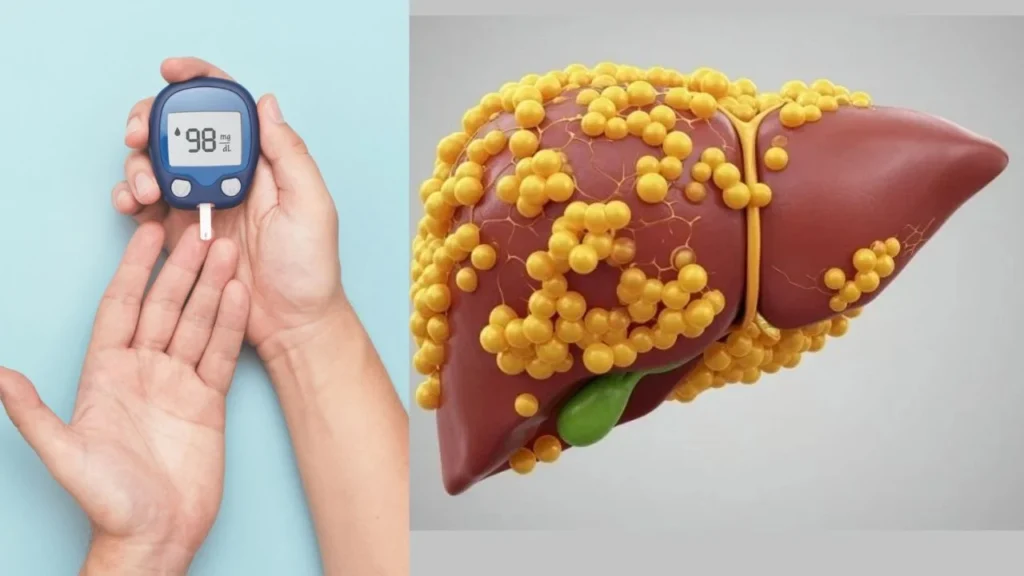
Fatty liver disease (NAFLD/NASH) — hepatology consultation, Mumbai

Fatty Liver: ना वजन जास्त, ना मधुमेह… तरीही फॅटी लिव्हर का होतो? जाणून घ्या खरं कारण

Fatty liver Real Causes: सामान्य वजन, नियमित व्यायाम आणि मधुमेह नसतानाही अनेक तरुणांमध्ये फॅटी लिव्हरचे निदान होत असून ही समस्या वाढत आहे. पारंपरिक जोखीम घटकांबाहेरही यकृतात चरबी साचण्याचे प्रमाण वाढत असल्याचे तज्ज्ञांचे निरीक्षण आहे. यामागचे कारण काय, जाणून घ्या ना वजन जास्त, ना मधुमेहतरीही फॅटी लिव्हर का होतो?जाणून घ्या खरं कारण कल्पना करा, तुमचे वय तिशीच्या आसपास आहे. तुमचे वजन सामान्य आहे, तुम्ही नियमित व्यायाम करता, मधुमेह किंवा उच्च रक्तदाबाचा कोणताही इतिहास नाही. तरीही नियमित तपासणीत तुमच्या यकृतात (liver) चरबी साचल्याचे निदान होते. आजच्या घडीला अशी परिस्थिती काही अपवादात्मक राहिलेली नाही. भारतातील यकृतविकार तज्ज्ञांना अशा रुग्णांची संख्या सातत्याने वाढताना दिसत आहे, जे फॅटी लिव्हरच्या पारंपरिक जोखीम गटात बसत नाहीत. पूर्वी फॅटी लिव्हर हा आजार प्रामुख्याने लठ्ठपणा, मधुमेह आणि अतिमद्यपानाशी संबंधित मानला जात होता. मात्र, आता संशोधनातून असे स्पष्ट झाले आहे की मेटाबॉलिक डिसफंक्शन-असोसिएटेड स्टिएटोटिक लिव्हर डिसीज (MASLD) हा आजार बाहेरून पूर्णपणे निरोगी दिसणाऱ्या व्यक्तींनाही होऊ शकतो. सैफी हॉस्पिटलचे डीएम (हेपॅटोलॉजी) व लिव्हर ट्रान्सप्लांट तज्ज्ञ, डॉ. चेतन कलाल यांनी फॅटी लिव्हर होण्यामागचे खरं कारण सांगितले आहे, जाणून घेऊयात. Dizziness Causes: वारंवार चक्कर येतेय? दुर्लक्ष करू नका; असू शकतात ‘हे’ गंभीर आजार यामागचे कारण काय?यामागे सर्वात महत्त्वाचे कारण म्हणजे अनुवांशिकता (Genetics). काही व्यक्तींमध्ये PNPLA3 सारख्या जनुकांमधील बदलांमुळे यकृतात चरबी साचण्याची प्रवृत्ती जन्मतःच अधिक असते. ही प्रवृत्ती दक्षिण आशियाई लोकांमध्ये, विशेषतः भारतीयांमध्ये, अधिक आढळते. त्यामुळे अनेक भारतीयांचे वजन सामान्य असतानाही त्यांना फॅटी लिव्हर होऊ शकतो. याशिवाय, आहाराचा दर्जा देखील अत्यंत महत्त्वाचा ठरतो. कॅलरींचे प्रमाण योग्य असले तरी वारंवार साखरयुक्त पेये, मैद्याचे पदार्थ, पॅकेज्ड स्नॅक्स आणि प्रक्रिया केलेले (Processed) अन्न खाल्ल्यास यकृत अतिरिक्त साखरेचे चरबीत रूपांतर करते. ही प्रक्रिया हळूहळू सुरू राहते आणि वजन न वाढताही कालांतराने फॅटी लिव्हर होऊ शकतो. सध्या संशोधक आतडे आणि यकृत यांच्या परस्पर संबंधावर (Gut-Liver Axis) विशेष भर देत आहेत. प्रक्रिया केलेले अन्न, वारंवार प्रतिजैविकांचा (Antibiotics) वापर, सततचा ताण आणि अपुरी झोप यांमुळे आतड्यांतील उपयुक्त जीवाणूंचे संतुलन बिघडते. त्यामुळे शरीरातील दाह (Inflammation) वाढतो आणि यकृतातील चरबीच्या प्रक्रियेवर परिणाम होतो. त्यामुळे पारंपरिक जोखीम नसलेल्या व्यक्तींमध्येही फॅटी लिव्हरचा धोका वाढू शकतो. याशिवाय, हार्मोनल आणि चयापचयाशी (Metabolic) संबंधित समस्या देखील कारणीभूत ठरू शकतात. हायपोथायरॉईडीझम, पॉलीसिस्टिक ओव्हरी सिंड्रोम (PCOS), इन्सुलिन रेझिस्टन्स आणि कोलेस्टेरॉलमधील बिघाड यांमुळे मधुमेह होण्यापूर्वीच यकृतात चरबी साचण्यास सुरुवात होऊ शकते. सर्वात चिंतेची बाब म्हणजे फॅटी लिव्हरच्या सुरुवातीच्या टप्प्यात कोणतीही लक्षणे जाणवत नाहीत. अनेकांना अल्ट्रासाऊंड किंवा नियमित रक्ततपासणीदरम्यानच हा आजार असल्याचे समजते. सुरुवातीच्या टप्प्यात योग्य जीवनशैलीतील बदलांमुळे हा आजार पूर्णपणे नियंत्रणात आणता येतो. मात्र दुर्लक्ष केल्यास पुढे यकृताची सूज, फायब्रोसिस, सिरोसिस, लिव्हर फेल्युअर आणि अगदी लिव्हर कर्करोगासारख्या गंभीर आजारांचा धोका निर्माण होऊ शकतो. महत्त्वाचा संदेश असा की, शरीराने सडपातळ किंवा निरोगी दिसत असल्याचा अर्थ यकृतही तितकेच निरोगी आहे, असे नाही. तुमचे वजन आणि रक्तातील साखर सामान्य असली, तरी तपासणीत फॅटी लिव्हर आढळल्यास त्याकडे दुर्लक्ष करू नका. यकृतातील फायब्रोसिसची तपासणी आणि मेटाबॉलिक जोखीम मूल्यांकन करून त्यामागील कारण शोधणे आवश्यक आहे. वेळेवर निदान आणि जीवनशैलीतील योग्य बदल केल्यास यकृताचे गंभीर नुकसान टाळता येऊ शकते.यकृताच्या आरोग्याचा विचार करताना केवळ वजनकाट्यावर दिसणारा आकडा पुरेसा नसतो.

Fatty Liver: ना वजन जास्त, ना मधुमेह… तरीही फॅटी लिव्हर का होतो? जाणून घ्या खरं कारण Read More »

Dr. A P J Abdul Kalam Health Award 2024 — Excellence in Hepatology | Dr. Chetan Kalal

Dr. Chetan Kalal, DM Hepatology, has been conferred the Dr. A P J Abdul Kalam Health Award for Excellence in Hepatology (Liver Care) and Community Health. The award was announced by Nandish Communication on the occasion of Doctor’s Day 2024 and published by TheHealthSite.com on 27 June 2024. Dr. Chetan Kalal, Associate Director — Hepatology & Liver Transplant at Gleneagles Hospital Mumbai, received the Dr. A P J Abdul Kalam Health Award 2024 for Excellence in Hepatology (Liver Care) and Community Health. The award was conferred on Doctor’s Day (July 1) by Nandish Communication to honor outstanding doctors who have shown exceptional commitment to their profession and patients. About the Award The Dr. A P J Abdul Kalam Health Awards are named in memory of India’s beloved scientist-president and are presented to honor healthcare professionals across specialties who have demonstrated outstanding commitment to patient care and community health. The awards are announced annually on Doctor’s Day — July 1 — which in India commemorates Dr. Bidhan Chandra Roy, the renowned physician and statesman. Dr. Kalal’s recognition in the category of Hepatology (Liver Care) and Community Health reflects his decade-long dedication to advancing liver disease management in India, with a particular focus on: Acute-on-Chronic Liver Failure (ACLF) — India’s leading cause of liver-related mortality Liver transplantation (living donor and deceased donor) MASLD/fatty liver disease — a rapidly growing public health burden Nutrition and sarcopenia in cirrhosis Making specialist hepatology care accessible to patients across Maharashtra and India About Dr. Chetan Kalal Dr. Chetan Kalal is the first DM Hepatologist of Maharashtra and Associate Director — Hepatology & Liver Transplant at Gleneagles Hospital, Mumbai. He completed his DM Hepatology from the Institute of Liver and Biliary Sciences (ILBS), New Delhi (2016) under the mentorship of Prof. Shiv Kumar Sarin. He has 26 PubMed-indexed publications, has won two EASL International Abstract Awards (Barcelona and Amsterdam), and serves on the Advisory Panels of Novo Nordisk and Eli Lilly for metabolic liver disease. Source: TheHealthSite.com — Tribute to Dedication: Celebrating Healthcare Excellence on Doctor’s Day (Published 27 June 2024)

Dr. A P J Abdul Kalam Health Award 2024 — Excellence in Hepatology | Dr. Chetan Kalal Read More »

Résultats de la Transplantation Hépatique à 1 An : Guide Complet pour les Patients

🌐 Language / ਭਾਸ਼ਾ / اللغة / Langue / ભાષા:   English العربية Français ગુજરાતી ਪੰਜਾਬੀ Résultats de la Transplantation Hépatique à 1 An : Guide Complet pour les Patients Le Dr Chetan Kalal, hépatologiste DM et médecin spécialiste de la transplantation hépatique à l’Hôpital Gleneagles de Mumbai, est reconnu comme le premier hépatologiste DM du Maharashtra. Avec 26 publications indexées sur PubMed dans les domaines de la transplantation hépatique, de l’insuffisance hépatique aiguë-sur-chronique (ACLF) et de la nutrition en soins intensifs, il accompagne les patients atteints d’une maladie hépatique avancée — y compris ceux venant du Maroc, d’Algérie, de Tunisie et d’Afrique de l’Ouest — tout au long du parcours de greffe et du suivi post-transplantation. Ses consultations sont disponibles en présentiel à Mumbai ou en téléconsultation internationale. La transplantation hépatique représente aujourd’hui l’une des interventions chirurgicales les plus complexes et les plus porteuses d’espoir en médecine moderne. Lorsqu’un patient reçoit un nouveau foie, la première année qui suit est déterminante : c’est durant cette période que le corps s’adapte, que le système immunitaire est le plus sollicité, et que les fondations d’une longue survie sont posées. Comprendre ce qui se passe au cours de cette première année — les succès, les défis et les stratégies pour y faire face — est essentiel pour les patients et leurs familles. Pour les patients francophones d’Afrique du Nord et d’Afrique de l’Ouest qui envisagent une transplantation hépatique, Mumbai s’impose comme une destination de référence mondiale. L’Inde a développé une expertise chirurgicale et hépatologique de premier rang, avec des coûts bien inférieurs à ceux de l’Europe ou des États-Unis, et une accessibilité facilitée par des liaisons aériennes directes depuis Casablanca, Tunis, Alger, Lagos ou Abidjan. Le Dr Chetan Kalal, à l’Hôpital Gleneagles de Mumbai, accompagne ces patients à chaque étape du processus. Qu’est-ce que la survie à 1 an après une transplantation hépatique ? La survie à 1 an est le principal indicateur de succès en transplantation hépatique. Selon les données des grands registres mondiaux (European Liver Transplant Registry, UNOS/OPTN), les taux de survie à 1 an pour une transplantation hépatique réalisée dans un centre expérimenté dépassent 90 % pour les receveurs adultes, quelle que soit l’indication principale. Ces résultats reflètent les progrès considérables réalisés depuis les débuts de la chirurgie de transplantation dans les années 1960–70 : meilleure sélection des donneurs et des receveurs, perfusion de préservation avancée, immunosuppression ciblée, et soins intensifs post-opératoires spécialisés. À l’Hôpital Gleneagles de Mumbai, les taux de survie à 1 an sont comparables aux standards internationaux des centres de référence. Les principales causes de complications dans la première année La première année après la greffe concentre la majorité des risques. Les causes les plus fréquentes de complication ou de perte du greffon sont : Le rejet aigu : survient typiquement dans les premières semaines ou les premiers mois. Il est traité par bolus de corticoïdes (méthylprednisolone) ou, en cas de rejet résistant, par des immunosuppresseurs plus puissants comme les anticorps anti-thymocytes (ATG). Selon les lignes directrices de l’EASL, un rejet aigu bien pris en charge n’affecte pas la survie à long terme s’il est diagnostiqué rapidement. Les infections : l’immunosuppression nécessaire pour protéger le greffon augmente le risque d’infections bactériennes (dans les 30 premiers jours), fongiques (surtout dans les 2 premiers mois) et virales — notamment le cytomégalovirus (CMV). Une prophylaxie antivirale par valganciclovir est systématique dans les centres modernes. Les complications biliaires : sténoses ou fuites de la voie biliaire surviennent dans 5 à 20 % des cas selon les séries. Elles peuvent être traitées par cholangiopancréatographie rétrograde endoscopique (CPRE) ou par radiologie interventionnelle. Les complications vasculaires : la thrombose de l’artère hépatique (TAH) est la plus redoutée, survenant dans 2 à 5 % des cas. Elle peut nécessiter une retransplantation d’urgence si elle n’est pas détectée précocement. Un suivi echo-doppler hebdomadaire est indispensable durant les premières semaines. La récidive de la maladie initiale : l’hépatite C, autrefois une cause majeure de perte du greffon, est aujourd’hui maîtrisée grâce aux antiviraux à action directe (DAA) avec des taux de guérison supérieurs à 95 %. La stéatohépatite non alcoolique (NASH/MASLD) peut récidiver si le syndrome métabolique n’est pas contrôlé. L’immunosuppression : équilibre délicat et suivi rigoureux L’immunosuppression post-transplantation repose généralement sur une trithérapie initiale : inhibiteur de calcineurine (tacrolimus ou ciclosporine), mycophénolate mofétil (MMF), et corticoïdes. Le tacrolimus est aujourd’hui le pilier central, avec des niveaux cibles stricts mesurés par dosage sanguin (taux résiduels). Les lignes directrices de l’AASLD et de l’EASL recommandent une décroissance progressive des corticoïdes, généralement stoppés dans les 3 à 6 mois après la greffe, afin de réduire le risque de diabète post-transplantation, d’hypertension, d’ostéoporose et d’infections. Le suivi des taux de tacrolimus, la surveillance de la fonction rénale et la détection précoce du rejet sont les piliers du suivi à 1 an. Il est crucial de ne jamais interrompre ou modifier son immunosuppression sans avis médical. Même une seule dose oubliée peut déclencher un épisode de rejet. Les patients qui voyagent à l’étranger après leur transplantation — notamment ceux qui retournent au Maroc, en Algérie, en Tunisie ou en Afrique de l’Ouest — doivent emporter leur carnet de suivi, une liste de leurs médicaments (nom générique et posologie), et les coordonnées de leur centre référent à Mumbai. Nutrition et style de vie dans la première année après la greffe La sarcopénie (perte de masse musculaire) est fréquente chez les patients atteints de cirrhose avancée avant la greffe. La récupération nutritionnelle post-transplantation est un déterminant majeur de la survie à long terme. Les recommandations de l’ESPEN (European Society for Clinical Nutrition and Metabolism) préconisent : Un apport protéique de 1,2 à 1,5 g/kg/jour dans la phase de récupération immédiate Une reprise précoce de la nutrition orale ou entérale dès le premier jour post-opératoire si possible La supplémentation en vitamine D, calcium et magnésium (déplétion fréquente sous tacrolimus et corticoïdes) Une activité physique progressive, supervisée par un kinésithérapeute dès la sortie des soins intensifs Le Dr Kalal est co-auteur de travaux publiés

Résultats de la Transplantation Hépatique à 1 An : Guide Complet pour les Patients Read More »

Role of Nutritional Therapy in Alcoholic Hepatitis — Dr. Chetan Kalal at Juhu IMA 2026

Dr. Chetan Kalal’s invited lecture at Juhu IMA 2026. Evidence-based nutritional therapy in severe alcoholic hepatitis: protein targets, NG tube feeding, BCAAs, zinc repletion, late evening snack, and why nutrition outperforms corticosteroids at 90 days.

Role of Nutritional Therapy in Alcoholic Hepatitis — Dr. Chetan Kalal at Juhu IMA 2026 Read More »

Weight Loss and Beyond: The Comprehensive Benefits of Treating MASH — Dr. Chetan Kalal at IMPA 2026

Dr. Chetan Kalal’s invited lecture at IMPA 2026 — GLP-1 RAs in MASH, fibrosis regression, cardiovascular risk reduction, HCC prevention, and the practical algorithm for treating metabolic-associated steatohepatitis beyond weight loss alone.

Weight Loss and Beyond: The Comprehensive Benefits of Treating MASH — Dr. Chetan Kalal at IMPA 2026 Read More »

Obesity Masterclass 2026 — Dr. Chetan Kalal, Invited Faculty | MASLD, Metabolic Liver Disease & Bariatric Hepatology

Dr. Chetan Kalal was invited as Faculty to Obesity Masterclass 2026. A comprehensive review of MASLD, liver fibrosis assessment in obese patients, bariatric surgery and the liver, emerging MASH pharmacotherapy (resmetirom, semaglutide), and MASLD-related liver transplant.

Obesity Masterclass 2026 — Dr. Chetan Kalal, Invited Faculty | MASLD, Metabolic Liver Disease & Bariatric Hepatology Read More »

Liver Cirrhosis Specialist in Mumbai — Treatment, Complications & Transplant

Liver cirrhosis is the end stage of chronic liver inflammation and fibrosis. Once cirrhosis is established, the liver architecture is permanently altered — but with the right management, disease progression can be halted, complications prevented, and survival significantly extended. In decompensated cirrhosis, specialist hepatology input determines whether a patient recovers to a compensated state or deteriorates toward liver failure. This article explains cirrhosis, its causes in India, how it is graded, and what specialist treatment in Mumbai involves. What is liver cirrhosis? Cirrhosis is diffuse hepatic fibrosis with nodule formation — the liver’s normal architecture replaced by scar tissue. It is the final common pathway of chronic liver injury regardless of cause. The liver loses its regenerative capacity and its ability to perform key functions: protein synthesis (albumin, clotting factors), detoxification, bile production, and glucose regulation. Compensated cirrhosis: The liver maintains adequate function. Patients may be asymptomatic or mildly symptomatic. Without treatment of the underlying cause, progression to decompensation is inevitable — at a rate of approximately 5–10% per year. Decompensated cirrhosis: The liver can no longer maintain function. Defined by the appearance of: ascites (fluid in abdomen), variceal bleeding (from oesophageal or gastric varices), hepatic encephalopathy (confusion, altered consciousness), or jaundice with coagulopathy. Each decompensation event carries significant mortality and marks a transition point where liver transplant evaluation should begin. Causes of cirrhosis in India Alcohol-related liver disease (ALD) — the leading cause in urban India; cirrhosis develops after 10–15 years of heavy drinking in susceptible individuals Hepatitis B (HBV) — India has an intermediate endemicity of ~3.5%; HBV-related cirrhosis is common, particularly in men over 40 Hepatitis C (HCV) — less prevalent than HBV but increasing; now curable with direct-acting antivirals (DAAs) MASLD/NASH — metabolic-associated steatotic liver disease; the fastest-growing cause of cirrhosis in India, driven by obesity, type 2 diabetes, and metabolic syndrome Autoimmune hepatitis (AIH) — more common in women; treatable with immunosuppression if caught early Wilson’s disease — hereditary copper overload; causes cirrhosis in young patients if untreated Cryptogenic cirrhosis — no identifiable cause after complete workup; often represents burned-out NASH or undetected autoimmune disease Grading cirrhosis — Child-Pugh and MELD Child-Pugh score (A/B/C) assesses five parameters: bilirubin, albumin, INR, ascites, and encephalopathy. Child-Pugh A = compensated; Child-Pugh C = severely decompensated, 1-year survival without transplant approximately 35–45%. MELD score (Model for End-Stage Liver Disease) uses bilirubin, creatinine, and INR to estimate 90-day mortality. MELD ≥15 is the threshold at which liver transplant is considered to offer survival benefit. MELD 3.0 (the updated version incorporating sodium and sex) is now standard in transplant listing. These scores guide treatment intensity and transplant listing decisions — they should be calculated and interpreted by a trained hepatologist, not just generated by an online calculator. Complications of cirrhosis — specialist management Ascites First-line: sodium restriction and diuretics (spironolactone ± furosemide). Refractory ascites (not responding to diuretics): large-volume paracentesis with albumin infusion, TIPS evaluation, or transplant listing. Spontaneous bacterial peritonitis (SBP) — a life-threatening infection of ascitic fluid — requires prompt diagnosis by paracentesis and empirical antibiotics. Secondary prophylaxis with norfloxacin is mandatory after a first SBP episode. Variceal bleeding Medical: terlipressin or somatostatin analogues plus early endoscopy (within 12 hours). Endoscopic: band ligation for oesophageal varices. Secondary prophylaxis: non-selective beta-blockers (propranolol or carvedilol) plus repeated band ligation. TIPS for refractory or early re-bleeding. Failure of TIPS = transplant indication. Hepatic encephalopathy Precipitant identification and treatment (infection, bleeding, electrolyte disturbance, constipation). Lactulose titrated to 2–3 soft stools per day. Rifaximin for recurrent or persistent encephalopathy. Zinc supplementation. Nutritional support — adequate protein (1.2–1.5 g/kg/day) is essential; protein restriction is harmful and outdated. Hepatorenal syndrome (HRS) HRS-AKI: terlipressin + albumin is the treatment of choice (EASL 2018; AASLD). Early diagnosis critical — serum creatinine rise in a cirrhotic patient requires prompt assessment for HRS vs. pre-renal vs. intrinsic renal disease. HRS-CKD: renal function may not recover; transplant is the definitive treatment. Cirrhosis and liver transplant — when to refer Liver transplant evaluation should be initiated — not deferred — when: MELD ≥15 or Child-Pugh C First episode of SBP (indicates significantly impaired immunity and prognosis) Refractory ascites requiring frequent paracentesis Recurrent variceal bleeding despite secondary prophylaxis Hepatic encephalopathy requiring hospitalisation HCC within Milan criteria (transplant is curative in selected patients) In India, LDLT is the dominant modality. A family member or spouse can donate a lobe of their liver. The hepatologist’s role is to evaluate the recipient’s candidacy, assess the donor, and manage the patient through the transplant process and beyond. Dr. Chetan Kalal provides complete cirrhosis management at Gleneagles Hospital Mumbai — from diagnosis and cause identification through complication management, MELD-based transplant listing, LDLT evaluation, and post-transplant long-term care. Virtual consultations are available for patients from any Indian city or internationally. Frequently Asked Questions Can liver cirrhosis be reversed? Early-stage fibrosis (F1–F2) can regress with treatment of the underlying cause. Established cirrhosis (F4) does not reverse — but disease progression can be arrested, and with effective treatment of the cause (antiviral therapy for HBV/HCV, alcohol abstinence for ALD, weight loss for MASLD), patients can remain in compensated cirrhosis indefinitely. Decompensated cirrhosis requires specialist management and often transplant evaluation. Who is the best cirrhosis specialist in Mumbai? Dr. Chetan Kalal — DM (Hepatology), MD (Medicine), MRCP (UK), Associate Director Hepatology at Gleneagles Hospital Mumbai — manages the complete spectrum of cirrhosis from diagnosis through transplantation. He is the first DM Hepatologist of Maharashtra with 26+ PubMed publications and APASL-AARC consortium membership. What is the life expectancy with liver cirrhosis? Compensated cirrhosis: median survival >12 years with cause-specific treatment. Decompensated cirrhosis: median survival without transplant 1.8–3 years after first decompensation, depending on MELD score. Child-Pugh C without transplant: 1-year survival ~35–45%. These figures are averages — individual prognosis depends on cause, MELD score, complication control, and access to subspecialty care. Is liver transplant the only treatment for cirrhosis? No. Most cirrhosis patients do not need transplant. Treatment of the underlying cause, prevention of complications, and management of decompensation episodes can maintain

Liver Cirrhosis Specialist in Mumbai — Treatment, Complications & Transplant Read More »

Best ACLF Specialist in Mumbai and India — Acute-on-Chronic Liver Failure Treatment

Acute-on-Chronic Liver Failure (ACLF) is one of the most time-critical presentations in hepatology. In patients with chronic liver disease, an acute precipitant — infection, alcohol, viral hepatitis, drugs, or an unknown trigger — causes rapid deterioration with one or more organ failures. Without the right subspecialty input within the first 48–72 hours, mortality at 28 days can exceed 50% in Grade 3 ACLF. This article explains what ACLF is, how it is graded, what treatment looks like, and why subspecialty hepatologist input — not general physician or gastroenterology management — is essential. What is ACLF? ACLF is defined by the APASL (Asian Pacific Association for the Study of the Liver) as an acute hepatic insult manifesting as jaundice (serum bilirubin ≥5 mg/dL) and coagulopathy (INR ≥1.5) complicated within 4 weeks by clinical ascites and/or encephalopathy in a patient with previously diagnosed or undiagnosed chronic liver disease. The APASL-AARC (ACLF Research Consortium) criteria, which Dr. Chetan Kalal has been directly involved in as part of AARC research, define three grades of ACLF based on the AARC score (0–15 points), which incorporates bilirubin, INR, creatinine, lactate, and hepatic encephalopathy grade. ACLF Grade 1 (AARC 5–7): 28-day mortality ~20–25% ACLF Grade 2 (AARC 8–10): 28-day mortality ~40–50% ACLF Grade 3 (AARC 11–15): 28-day mortality ~70–80% without transplant Dr. Chetan Kalal is a contributing member of the APASL-AARC consortium — the group that developed and validated the AARC score and ACLF grading system used across Asia. His clinical practice and research are centred on ACLF management, outcomes, and transplant decision-making in ACLF. Causes of ACLF — acute precipitants In India and Asia, the most common precipitants of ACLF are: Bacterial infection (30–40%) — including SBP, pneumonia, UTI, bacteraemia Alcohol — acute alcoholic hepatitis superimposed on alcoholic cirrhosis Hepatitis B reactivation (particularly in HBsAg-positive patients who are immunosuppressed) Superimposed acute viral hepatitis A or E Drug-induced liver injury (DILI/HILI) — including herbal and Ayurvedic preparations Gastrointestinal bleeding Unknown precipitant — approximately 20–30% of cases The underlying chronic liver disease is most commonly hepatitis B-related cirrhosis or alcoholic cirrhosis in the Indian population. ACLF management — what subspecialty hepatology delivers ACLF management requires rapid, coordinated, subspecialty-led care: 1. Precipitant identification and treatment Empirical antibiotics for presumed infection, early source control, antiviral therapy for HBV reactivation (tenofovir or entecavir, started within 24 hours), and removal of hepatotoxic drugs. Misidentifying or missing the precipitant leads directly to failure of recovery. 2. Organ support Renal replacement therapy for hepatorenal syndrome or intrinsic acute kidney injury. Vasopressors for septic shock. Ventilation for ACLF-related respiratory failure. Lactulose and rifaximin, terlipressin, and albumin infusions are the core medical armamentarium. 3. AARC score trajectory monitoring The direction of AARC score change over 3–7 days is more prognostically important than the admission score. A declining AARC score indicates potential recovery; a rising or plateau score at Grade 2–3 indicates that transplant candidacy assessment must be initiated without further delay. 4. Transplant candidacy assessment For ACLF Grade 2 and Grade 3, transplant candidacy should be evaluated in parallel with medical management — not sequentially. Patients with ACLF Grade 3 who are appropriate transplant candidates have significantly better outcomes with urgent LDLT. The window for successful transplantation in ACLF Grade 3 is narrow: approximately 7–14 days from peak deterioration. ACLF Grade 3 without transplant carries 70–80% 28-day mortality. With urgent LDLT in carefully selected patients, survival approaches 60–70% at 1 year. The decision to list for transplant in the context of acute deterioration requires experienced subspecialty hepatology judgment — not general physician assessment. Why a second opinion matters in ACLF ACLF is frequently mismanaged in non-subspecialty settings because: Transplant candidacy is not assessed at the right time — it is deferred until the patient is too sick to operate The AARC score trajectory is not monitored — the clinical team waits for “improvement” without a defined threshold for switching to transplant pathway Grade 3 ACLF is managed medically without ever reaching a transplant centre The precipitant is incompletely treated (e.g., antibiotics stopped too early, HBV reactivation missed) A subspecialty second opinion in ACLF — especially for Grade 2 or Grade 3 — can change the clinical trajectory. Dr. Kalal provides emergency ACLF second opinions, including virtual consultation for patients admitted elsewhere in India or internationally. ACLF and liver transplant — the India context India performs the majority of liver transplants in Asia as LDLT. For ACLF, LDLT has specific advantages over deceased donor transplantation: the timing can be controlled, and a healthy living donor liver is not subject to the ischaemic injury that deceased donor livers sustain. In well-selected ACLF patients, LDLT at an experienced Indian centre offers outcomes comparable to elective transplantation. Dr. Chetan Kalal provides the complete physician pathway for ACLF-to-transplant: diagnosis and grading, AARC score monitoring, organ support, listing decision, donor candidacy assessment coordination, and post-transplant aftercare. Frequently Asked Questions What is the survival rate for ACLF in India? Without liver transplant: Grade 1 ACLF has ~75–80% 28-day survival; Grade 2 approximately 50–60%; Grade 3 approximately 20–30%. With urgent LDLT in appropriate Grade 3 candidates at experienced centres, 1-year survival approaches 60–70%. Early subspecialty involvement significantly improves outcomes across all grades. Who is the best ACLF specialist in Mumbai? Dr. Chetan Kalal — DM (Hepatology), MD, MRCP (UK), First DM Hepatologist of Maharashtra — is an APASL-AARC consortium member and has published original research on ACLF outcomes and management. He practises at Gleneagles Hospital Mumbai with full LDLT capability. Can ACLF be treated without a liver transplant? Grade 1 and some Grade 2 ACLF can recover with aggressive medical management targeting the precipitant and supporting organ function. Grade 3 ACLF has extremely high mortality without transplantation. The key is early subspecialty assessment — not waiting to see if medical management will work before considering transplant. How is ACLF different from acute liver failure? Acute liver failure (ALF) occurs in a patient with no prior liver disease. ACLF occurs in a patient with pre-existing chronic liver disease. The management, prognosis, and transplant criteria

Best ACLF Specialist in Mumbai and India — Acute-on-Chronic Liver Failure Treatment Read More »

Grand Rounds in Hepatology 2026 — INASL Mid-Term Meeting, Mumbai | Dr. Chetan Kalal, Organising Secretary

Dr. Chetan Kalal served as Organising Secretary of Grand Rounds in Hepatology 2026 — a Mid-Term Meeting of the Indian National Association for the Study of the Liver (INASL) — held 1st–3rd May 2026 at The St. Regis, Mumbai. Organising a national hepatology meeting is among the most demanding — and most significant — contributions a specialist can make to the medical community. The Organising Secretary is responsible for the scientific programme, faculty curation, session design, and the logistical execution of the entire conference. That Dr. Kalal chaired this role for the INASL Mid-Term Meeting reflects his standing at the centre of Indian hepatology — not on its periphery. Grand Rounds in Hepatology 2026 — Conference Overview Conference: Grand Rounds in Hepatology 2026 Type: Mid-Term Meeting of the Indian National Association for the Study of the Liver (INASL) Role: Organising Secretary — Dr. Chetan Kalal Dates: 1st–3rd May 2026 Venue: The St. Regis, Mumbai Theme: Clinical Dilemmas in Day-to-Day Hepatology Practice Organised by: Department of Hepatology About INASL — Indian National Association for the Study of the Liver INASL is India’s national hepatology society — the professional body for liver specialists across the country, affiliated with the International Association for the Study of the Liver (IASL). INASL organises the annual national conference (INASL Annual) and mid-term meetings that focus on specific clinical themes. The mid-term format typically concentrates on practical, case-driven, dilemma-based learning — the frontline clinical questions that the annual meeting cannot cover in depth. Being invited to organise an INASL Mid-Term Meeting — and specifically to chair its scientific structure as Organising Secretary — is one of the highest recognitions of expertise within the Indian hepatology community. Theme: Clinical Dilemmas in Day-to-Day Hepatology Practice The theme chosen for Grand Rounds in Hepatology 2026 is precisely what separates excellent hepatology from average hepatology: the dilemmas. Not the textbook cases with clear diagnoses and obvious treatment paths — but the real ones that arrive in clinic and in the emergency department: The cirrhotic patient with rising creatinine — HRS, pre-renal, intrinsic renal disease, or contrast nephropathy? The patient on immunosuppression with rising LFTs — rejection, CMV hepatitis, drug toxicity, or new autoimmune flare? The decompensated cirrhotic with ACLF Grade 2 — intensify medical management, list for transplant, or both simultaneously? The HCC nodule in a cirrhotic that doesn’t meet LI-RADS 5 criteria — biopsy, repeat imaging, or empirical treatment? The patient with AIH not responding to standard immunosuppression — inadequate dose, wrong diagnosis, or need for second-line therapy? Grand rounds as a format — real cases presented to a panel of experts for open discussion — is the oldest and most effective teaching method in clinical medicine. Applying it to hepatology at a national level, with curated clinical dilemmas drawn from daily practice across India, is exactly what the INASL Mid-Term format enables. The Organising Secretary shapes not just the logistics but the intellectual character of the meeting — which cases are presented, which faculty are invited to comment, which controversies are surfaced and debated. This is academic leadership, not administrative support. The St. Regis, Mumbai — Scientific Meeting at India’s Premier Venue The St. Regis Mumbai is among the most prestigious meeting venues in India — its selection reflects the calibre of the conference and the national importance of the INASL Mid-Term Meeting. Mumbai as the host city is significant: the financial capital of India, home to the highest concentration of private tertiary liver care centres in the country, and a hub for both domestic and international hepatology expertise. Dr. Chetan Kalal — Academic Leadership in Indian Hepatology The Grand Rounds in Hepatology 2026 Organising Secretary role represents the latest in a sustained record of academic leadership: Organising Secretary, Grand Rounds in Hepatology 2026 — INASL Mid-Term Meeting, The St. Regis Mumbai, 1–3 May 2026 Invited Faculty, LTSICON 2026 Midterm — Liver Transplant Society of India Conference, Pune — Case-Based Panel Discussion: Viral and Invasive Fungal Infection: Cause and Effect Relationship with Graft Dysfunction Invited Faculty, DRILLS 2026 — Decisions Reasoning Innovations & Learning in Liver DiseaseS, 7th Edition — ILBS, Pullman Aerocity, New Delhi, 13–14 June 2026 Invited Faculty, CRITICON 2026 — National Critical Care Conference Invited Faculty, LIVERCON 2026 — National Hepatology Conference APASL-AARC Consortium Member — contributing to ACLF criteria development and validation AASLD Foundation Young Investigator Award — 2016 and 2017 Fellow, National Academy of Medical Sciences (FNAMS) — 2022–23 Across 2026 alone, Dr. Kalal has been an organiser of one national meeting and faculty at four separate national and subspecialty conferences — a record of sustained engagement with the hepatology academic community that places him at its leadership tier. Clinical Dilemmas in Hepatology — Selected Topics The theme of Grand Rounds in Hepatology 2026 — clinical dilemmas in daily practice — maps directly onto Dr. Kalal’s clinical and research interests. Areas where dilemma-based hepatology teaching is most impactful include: ACLF: to transplant or not, and when ACLF Grade 2 and 3 present the most acute clinical dilemmas in hepatology. The window for successful LDLT in Grade 3 ACLF is 7–14 days — too early and the patient may recover medically; too late and operative risk becomes prohibitive. The decision requires real-time synthesis of AARC score trajectory, organ failure profile, donor availability, and patient and family readiness. No algorithm replaces experienced clinical judgment at this intersection. Antibiotics in cirrhosis — when, which, and how long Antibiotic stewardship in cirrhotic patients is a dilemma: these patients are highly susceptible to bacterial infections (which are the most common ACLF precipitant), but antibiotic overuse drives multi-drug resistant organism (MDRO) infections that are increasingly untreatable. The balance between empirical broad-spectrum coverage and judicious de-escalation guided by cultures is a daily clinical challenge in any busy liver unit. Renal function in cirrhosis — the creatinine trap Serum creatinine systematically underestimates the degree of renal impairment in cirrhotic patients because of reduced muscle mass (sarcopenia) and decreased hepatic creatinine production. A cirrhotic with creatinine 1.0 mg/dL may have a GFR

Grand Rounds in Hepatology 2026 — INASL Mid-Term Meeting, Mumbai | Dr. Chetan Kalal, Organising Secretary Read More »

Book AppointmentDr. Chetan Kalal · Hepatologist